Single Particle Cryo-TEM School
Single Particle Cryo-TEM School
This course provides an introduction to the single particle cryo-TEM pipeline for solving high resolution structures of macromolecular complexes. The 4 day course focuses on providing hands on training for sample preparation by plunge freezing (Vitrobot Mk 4, Leica EM GP) and loading into Titan Krios microscopes, as well as demonstrations of high pressure freezing and non-blotting (spraying) freezing devices. With small group (3 participants) teaching, delegates will get a day on the Titan Krios microscopes to become familiar with imaging/data collection using a Titan Krios microscope with EPU software. Course participants have the option to bring their own samples to prepare grids, and may have a limited opportunity to image and collect data during the course. We welcome course participants with a range of experience and backgrounds, including from academia and industry.
This course is now full, however if you would like to be added to the waiting list please email Karina@rms.org.uk
Outline Schedule
This is the outline schedule for the course. A more detailed programme will be available soon.
The course will begin on Monday 10 December at 09.00 and finish on Thursday 13 December at 14.30. You can bring your own samples to work on during the course.
Day1
Lectures on:
- Building projects up for Cryo-EM
- Cryo-EM Sample Prep
- Emerging Technologies for Sample Prep
- Microscope Hardware
- Single Particle TEM/Low Dose Imaging
- Electron Tomography
Day 2-4
Five half day Practical Sessions including two sessions on Titan Krios microscopes, and vitrification using the Vitrobot Mk 4, EM GP, and other freezing instruments (non blotting devices, high-pressure freezing)
Day 4
Half day of lectures on Image Processing
Organiser and Speaker List
Organiser
-

Dr Rebecca Thompson
Electron Microscopy Deputy Chair
University of Leeds
Rebecca is Facility Manager and senior cryo-electron microscopy (EM) support scientist at the Astbury Biostructure Laboratory, University of Leeds. Her research interests include imaging a broad range of biological specimens, from whole cells to macromolecular complexes, to high resolution using cryo-EM, and integrating data from EM with other microscopy techniques.
Confirmed Speakers
-
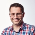
Mr Alan Boswell
Thermo Fisher
Alan is an experienced senior field service engineer for Thermo Fisher Scientific, formerly FEI. With over 30 years’ experience working with transmission electron microscopes, Alan has specialised in installing and maintaining Titan Krios systems since their introduction into the market ten years ago. He mentors trainee engineers using his teaching skills, and is regularly asked to contribute to product development and projects improving the customer experience. He has recently accepted the challenge of service manager for Thermo Fisher TEM life science within the UK.
-
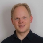
Dr Dan Clare
Diamond Light Source
I am Senior Beamline Scientist at the electron-Bioimaging Centre (eBIC) the UK’s national cryo-electron microscopy (cryo-EM) facility based at Diamond light source. My role at eBIC involves co-managing the cryo-EM facility and the international user program that we offer on our four Titan Krios microscopes. Before eBIC I was a senior post-doc in the laboratory of professor Helen saibil at Birkbeck College in London. I am a structural biologist by training and have had a longstanding interest in using cryo-EM and single particle analysis to look at how molecular chaperones function. I also have an active interest in cryo-electron tomography as a way of structurally charactering more complicated systems and currently have a research project looking at the bacterial infection of mammalian cells.
-
Dr Juan Fontana
University of Leeds
Juan Fontana has over 13 years of experience in structural virology and electron microscopy (EM). During his PhD (CNB-CSIC, Madrid, Spain), he characterised the replication complexes of Bunyamwera and Rubella viruses. This work allowed him to elucidate the mechanisms by which viral macromolecular complexes interact with cellular components to create the architectures of viral factories and to propose novel working models for each replication complex. As a post-doc (LSBR, NIH, Maryland, USA), he worked on Herpes Simplex Virus (HSV) & Influenza entry and on HIV & HSV morphogenesis. These projects included, 1) Elucidating the mechanism of Herpes Simplex Virus (HSV) neutralization by antibodies directed at the fusion domains of HSV’s fusion protein. 2) Unraveling structural intermediates of Influenza Virus fusion protein and M1 matrix protein prior to viral fusion. 3) Describing the two-step fusion process of the retrovirus Avian Sarcoma/Leukosis, in which receptor binding necessarily precedes low-pH activation of the fusion protein. 4) Studying the effects of several drugs and mutations that affect HIV maturation, an essential process for viral infectivity that involves proteolysis of precursor proteins by the viral protease and assembly of a conical core. Overall these projects gave Juan an in-depth knowledge of cell culture, manipulation of viruses, fluorescence & confocal microscopy, and cellular EM; and an expert knowledge of cryo-EM, including sample preparation, data acquisition, high-resolution single-particle reconstruction & atomic modeling and cryo-electron tomography & subtomogram averaging.On April 2016, Juan started a tenure-track academic fellowship at the University of Leeds. His lab uses cryo-EM, combined with other structural approaches, to characterise macromolecular assemblies, and viruses that are a current public health risk, like HIV, Influenza, herpesviruses and bunyaviruses.
-
Dr Emma Hesketh
University of Leeds
Emma joined the university of Leeds in 2015 as a postdoctoral researcher in Neil Ranson's group. During this time her research interests focused on the structure of viruses using cryo-electron microscopy to preform single particle 3D reconstructions to high (<3.5Å) resolution. Emma’s role now is as a cryo-EM Support Scientist in the Astbury Biostructure Laboratory and is involved in a broad range of research questions all using high resolution electron microscopy.
-
Dr Matthew Iadanza
University of Leeds
Matt completed his Ph.D. in the lab of Dr. Tamir Gonen at the University of Washington in Seattle, where he worked developing microcrystal electron diffraction (microED). He is currently a senior research fellow in the labs of Sheena Radford and Neil Ranson at the University of Leeds, where he uses single-particle and helical reconstruction cryoEM techniques to study the structure of amyloid fibrils and the folding and insertion of proteins into the outer membrane of Gram-negative bacteria. -

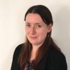
Rachel Johnson
University of Leeds
Rachel is a final year PhD student at the University of Leeds working under the supervision of Stephen Muench and Colin Fishwick. Her PhD project uses cryo-EM to aid structure-based drug design and focuses on determining the structure of membrane proteins which could act as anti-parasitic therapeutic targets -
Mr David Kleb
University of Leeds
David is a student on the 4-year Wellcome trust PhD programme 'The Molecular Basis of Biological Mechanisms' at the Astbury Centre, University of Leeds. He is supervised by Stephen Muench and Frank Sobott and currently in his second year. His project is focused on developing a rapid grid preparation technique for cryo-EM which he aims to apply to time resolved structural studies.
-
Dr Dimitrios Kontziampasis
University of Leeds
Dr Dimitrios Kontziampasis is currently working in the Astbury Centre for Structural Molecular Biology, on the development of methodologies that can trap conformational states in the μs-ms timeframe for time resolved single particle cryo-Electron Microscopy. His main interests revolve around inter/crossdisciplinary and applied research, with a focus on the surface modification of materials for biological, energy and several other applications, and their characterisation. -
Dr Dan Maskell
University of Leeds
I completed my PhD at UCL working on biochemical and structural characterisation of protein complexes involved in kinetochore formation. I continued my research interests with postdoctoral studies into structural and functional characterisation of nucleoprotein complexes involved in retroviral integration using single particle EM and other biochemical techniques. I took another postdoctoral position at Leeds Where I worked on structural characterisation of viral genomes and their association with capsid proteins. Within Leeds I changed roles to become a support scientist at the Astbury Biostructure Laboratory, working with scientists to facilitate their use of EM to solve interesting biological problems. -
Dr Stephen Muench
University of Leeds
Dr Stephen Muench is a lecturer in membrane proteins at the Univeristy of Leeds. Having trained in X-ray crystallography, he moved to the field of electron microscopy in 2006. His main interests are in using electron microscopy, primarily single particle cryo-EM, to resolve the structure and mechanism of membrane proteins and to use this to underpin inhibitor design -
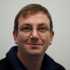
Dr Colin Palmer
Science and Technology Facilities Council
Colin Palmer is a structural biologist at STFC, working on CCP-EM, the Collaborative Computational Project for Electron cryo-Microscopy. After a PhD on electron tomography and more recent work in software engineering, his current role combines both these interests by focussing on computational methods for cryo-EM
-
Dr Neil Ranson
University of Leeds
-
Dr Christos Savva
University of Leicester
I am the facility manager at the Midlands Regional Cryo-EM Facility based at the University of Leicester. Prior to this I was a research assistant at the MRC Laboratory of Molecular Biology in Cambridge and an associate research scientist at Texas A&M University Microscopy and Imaging Center. I am a structural biologist with interest in the structure-function relationships of pore-forming proteins made by micro-organisms and bacteriophages. -
Dr Charlotte Scarff
University of Leeds
Dr Scarff is a Wellcome Trust ISSF Early Career Research Fellow at the University of Leeds. Her research interests lie in combining mass spectrometry and cryo-electron microscopy approaches to understand heterogeneity in macromolecular systems.
Further Information and Venue
This course will take place at the University of Leeds.
Registration fees and accommodation
Registration fees for this 4 day course are as follows:
RMS Member rate £1100
Non-member rate £1200
Accommodation package £264
Accommodation can be booked during the registration process. Accommodation has been reserved at the Ibis Hotel Leeds Centre Marlborough Street.
The accommodation package includes 4 nights bed and breakfast accommodation and transportation between the hotel and the course each day.
Sponsors
Confirmed Sponsors
-

Agar Scientific
Agar Scientific. Microscopy, Lab and Medical supplies at your fingertips
A leading international supplier of scientific instruments and accessories for over 40 years, Agar Scientific specialises in consumables and equipment supporting all forms of microscopy. Agar Scientific provides a fast, efficient online and offline ordering service, allowing customers to purchase with ease from our extensive range of laboratory and medical supplies and equipment. Our technical support staff has a wealth of experience in specimen preparation and microscopy techniques.
Our online catalogue of microscopy products includes:
TEM Grids - Apertures - Calibration standards - SEM stubs - Specimen preparation equipment - Filaments - Tweezers - Slides & cover glasses - Vacuum coating - Fixatives and resins - EM support films - Magnifiers - Specimen storageWe welcome enquiries regarding the use of any of our products, along with any suggestions for additions to our range. Orders ship from our UK headquarters in Stansted, Essex, and through our established network of agents and distributors who provide technical and applications support worldwide.
ISO9001:2008
Agar Scientific maintains a documented quality plan specifying our manufacturing and distribution processes and is approved by SGS to ISO9001:2008.Find out more about Agar Scientific
Web: www.agarscientific.com LinkedIn: Agar Scientific Twitter: @agarscientific -

Gatan
Gatan is the industry leader in the research, development and manufacturing of SEM and TEM products.
For TEM, Gatan provides an unrivalled range of high performance digital imaging and analytical systems, especially EELS spectrometers and energy filters (EFTEM). For SEM, Gatan are world leaders in cathodoluminescence (CL) technology and complement this with cooling stages and EBIC products.
Gatan is striving for continuous product innovations to increase users' productivity and make electron microscopes more effective and powerful tools. Visit the website for complete information on Gatan's extensive range of products for electron microscopy.
Find out more about Gatan
www.gatan.com -

Leica Microsystems
Leica Microsystems develops and manufactures microscopes and scientific instruments for the analysis of microstructures and nanostructures. Ever since the company started as a family business in the nineteenth century, its instruments have been widely recognized for their optical precision and innovative technology. It is one of the market leaders in compound and stereo microscopy, digital microscopy, confocal laser scanning microscopy with related imaging systems, electron microscopy sample preparation, and surgical microscopes.
Leica Microsystems has seven major plants and product development sites around the world. The company is represented in over 100 countries, has sales and service organizations in 20 countries, and an international network of distribution partners. Its headquarters are located in Wetzlar, Germany.
Find out more about Leica Microsystems
www.leica-microsystems.com -

Thermo Fisher Scientific
At Thermo Fisher Scientific, we are committed to accelerating your science by providing a comprehensive suite of solutions for the analysis of cells and their function. Behind this commitment is an incredible team of scientists developing and supporting our innovative instrumentation and products such as the Invitrogen™ Attune™ NxT Flow Cytometer, Invitrogen™ eBioscience™ flow cytometry antibodies, and Invitrogen™ functional reagents. Our flagship products are designed to deliver high-performance results and save you time in the lab. From our extensive flow cytometry cell health reagent portfolio and everyday standards and controls to our Invitrogen™ eBioscience™ Super Bright antibody conjugates, let us help you find unique ways to support discovery of new biological insights.
Find out more about Thermo Fisher Scientific
www.thermofisher.com -

TTP Labtech