Browse through articles previously published in infocus Magazine. If you are an RMS member you can view any article. Non members will be able to read articles over a year old.
Peroxisomes are essential organelles found in all eukaryotic cells. They are membrane bound compartments that play a critical role in lipid metabolism and detoxifying toxic products, such as hydrogen peroxide.
Atomic force microscopy (AFM) is a powerful technique often used to study surfaces at the nanoscale, helping researchers to better understand the structure and properties of materials.
When financial barriers stand between talented students and research experience, potential is lost. The BBSRC-funded BioImagingUK and UK Physics of Life Summer Studentships programme set out to change that — and the results speak volumes about what's possible when we remove obstacles to opportunity.
infocus is delighted to feature an interview with Professor Kang-Nee Ting, who was recently announced as winner of the 2025 RMS Chris Hawes Outreach and Education
Award.
A major outreach and education milestone was recently passed, as the total number of primary school-aged children to have benefitted from the RMS Microscope Activity Kits (MAKs) - since the scheme was established 15 years ago - went beyond 200,000.
We are the Electron Microscopy Scientific Technology Platform team from the Francis Crick Institute in London, and we share our experience of developing and delivering our Science Ambassador programme ‘Picturing Science in Schools’ to two local primary schools in the London borough of Camden.
As cancer continues to have a devastating effect on so many of us, it is only natural that people want to be informed about the activities of the world’s leading cancer research organisation: Cancer Research UK (CRUK).
Metastasis is the leading cause of cancer mortality. Neutrophils, a type of immune cell, play important roles in cancer with both potent anti- and pro-tumour activities.
As an international society, the RMS has always sought to strengthen links with other organisations dedicated to furthering the science of microscopy around the world.
Personally, I like recording videos of interesting or unusual things under the microscope to send to friends and family, or to include in publications.
The RMS is very pleased to announce the winners of its latest awards, recognising scientific achievement, contributions to education and outreach, technical support and services to the Society.
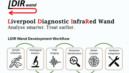
We are moving into a new era in microscopy technology development in which the microscopy technologies, in addition to the imaging function they have always provided, increasingly provide us with additional predictive and prognostic information which would be impossible to derive by any other method.
Formed in 1988, the Flow Cytometry Section is made up of a cross section of cytometrists representing research, clinical and industrial aspects of flow cytometry.
Our recently acquired collection of antique microscope slides includes some in the category “marine zoology”. The earliest members of our society – mainly wealthy, professional gentlemen – were keen to explore the new world that was opening up under their microscopes, and having neither the time nor skill to make their own, were avid collectors of slides made and sold by well-known mounters.
The scientific community increasingly recognises that laboratory practices can significantly contribute to the environmental footprint. At AstraZeneca, our ambitious sustainability strategy extends beyond corporate policies to fundamentally transform how we conduct research across our organisation and supply chain.