The program is open to UK-based technicians, research assistants, post-doctoral fellows, PhD students nearing the end of their studies, or researchers in equivalent positions in their careers. Selected applicants will have the chance to shadow a technical specialist for up to a week at a different institution's facility. This experience will provide them with insights into the daily operations of the facility, including operating various imaging modalities and software tools, managing open user access, ensuring quality management, and handling image data storage and analysis.
The program intends to facilitate the exchange of experiences and ideas between the hosting facilities and their guests. It also aims to encourage networking and foster possible future collaborations between imaging facilities and research groups.
If you are interested in participating in the program, make sure to review the specific application requirements and deadlines. Feel free to email Georgina Fletcher (Georgina@rms.org.uk) if you have any further questions or need more information.
Thank you to the Technician Commitment and the Technical Specialist Network for supporting the 2023 pilot of this scheme.
Applications Now Open
From 1 June - 31 August
Eligibility: The program is open to UK- or Ireland-based technicians, research assistants, post-doctoral fellows, PhD students nearing the end of their studies, or researchers in equivalent positions in their careers.
Program Details: The program offers the opportunity to shadow a technical specialist for up to a week at an imaging or flow cytometry core facility in the UK. Successful applicants will observe and learn about the technical job role, including operating imaging modalities and software tools, open user access, quality management, and image data analysis.
Application Requirements: Applications will reopen in June 2026. To apply, you need to complete this application form by 31 August 2026, which should include a letter of support from your current manager/supervisor and a letter from your desired host facility. The host facility's letter should confirm their agreement to the visit and briefly outline a plan for your time at the facility.
Assessment and Selection: Applications will be evaluated based on their fit with the Technician Commitment values and the potential impact on your career. The selection outcomes will be communicated in mid-September, and if selected, you will have the opportunity to shadow a technical specialist anytime up to November 30, 2026.
Expenses: If your application is successful, travel and lodging expenses of up to £500 will be covered.
Feedback and Publication: Both the guest and host will be required to complete a brief feedback form. The results will be compiled and written up for the RMS magazine, infocus.
To apply for the Technical Specialist Job Shadowing Scheme, you need to access the application form (https://forms.gle/sG6oaZZ7Puki25uB6). Fill out the form and make sure to include the required letters of support and agreement from your supervisor and desired host facility. Ensure that your application aligns with the Technician Commitment values and emphasises the potential impact on your career.
If you have any further questions or need additional assistance, please email Georgina Fletcher.
Technical Specialist Job Shadowing Organisers

Georgina Fletcher
Georgina is the Project Officer for the community network BioImagingUK, an open organisation of UK scientists that develop, use, or administer imaging solutions for life science research.
Contact Georgina for BioImagingUK enquiries.
georgina@rms.org.uk
+44 (0) 1865 254777


Alex Sossick
Alex is Head of Science Innovation Platforms at the Natural History Museum. He has a strong background in light microscopy, previously leading the Advanced Imaging Facility at the Gurdon Institute, University of Cambridge, which includes a variety of microscopy techniques including confocal, high throughput and deconvolution. He is keen to raise skills and access to technology and runs various courses.
Testimonials
I spent a week working at the Crick Advanced Light Microscopy STP shadowing Dr Dave Barry, one of the in house image analysts. During the week, I had the opportunity to participate in bioimage analysis user meetings, to meet imaging scientists and other image analysts working with light and electron microscopy data, and to join a half-day training on the Open Microscopy Environment (OMERO).
I learnt a lot in terms of how to give use support for bioimage analysis and in general on how a core service is run. Thanks to the shadowing project and other initiatives of the RMS DAIM committee, I will be working with my host and other committee members delivering a 2-day image analysis training in Ireland and some courses in London. Moreover, we are working on a paper on best practices for acquiring data for successful image analysis.
As part of the Technical Specialist Job Shadowing Scheme I visited the Experimental Techniques Centre at Brunel University London. The Experimental Techniques Centre has a strong background in material characterisation, and through this scheme I was able to spend time with all of the Scientific Officers that work there. Some of the technologies explored included Electron Microscopy (SEM and TEM), X-Ray Analysis (XRD and XRF) and Optical Spectroscopy (UV-vis, FTIR and Raman). During my time at the Experimental Techniques Centre I also gained valuable insights in to how the facility operates, how the finances are managed as well as how the quality of the work is managed in relation to UKAS accreditation. Experiences were shared and possible collaborative opportunities were discussed.
Following on from this visit, and the initial article within the RMS magazine, infocus, the primary goal is to collaborate with the Experimental Techniques Centre to build out a grant proposal that effectively utilises our respective expertise. We have already discussed potential ideas and hope to translate this to an upcoming funding call. I would say the most impactful outcome of this visit would be to subsequently forge a collaborative relationship between the materials groups at the University of Surrey and Brunel University London. It would also be great to use this as a spring board for championing technical expertise.
I visited the Bioscience Technology Facility at the University of York for 2 days. Through my visit I was able to discuss how better manage and integrate different platforms under the same technology facility - mainly flow cytometry and microscopy. My host was great, helping me with how to better manage my team and to consolidate and integrate my own experience in my facility.
I will be able to better integrate these technologies in my facility, where I will be able to get better working models to work with. For the future, we are looking for some collaborative work between University of York and University of Glasgow.
I visited the Advanced Imaging Resource at the Institute of Genetics and Cancer in Edinburgh. I mostly shadowed their bioimage analyst Laura Murphy, as well as other people who work with the facility.
The shadowing visit made me understand better the day-to-day work of a bioimage analyst. For example, I took part in a drop-in session where researchers brought images to get advice on analysis and quantification. I got in touch with several people who might be able to advise me in my future job of software specialist in data management (starting January 2025), and who can advise me in the future to find a bioimage analyst job. I wrote a short summary of the visit in blog format for the institute's website: https://blogs.ed.ac.uk/institute-genetics-cancer/2024/10/08/advanced-imaging-resource-job-shadowing-visit/
I spent a week at the Central Cell Imaging facility at Liverpool university. I shadowed the manager and a technician during the week, joining their pilot and induction training sessions. The visit included a tour of the facilities and meetings with relevant staff.
The visit was hugely beneficial as it allowed me to see how a central imaging facility is run and managed, as well as learning how to use various microscopes that I have not previously had access to. It greatly widened my knowledge on new instruments and also strengthened my understanding and expertise of my own equipment.
I have discussed the visit with my colleagues who may potentially want to use the facility and form collaborations on similar studies.
I have kindly been informed that I can keep in touch with my hosts for any issues or further questions I may have with my equipment so I have gained some useful support contacts for the future.
The visit was also very useful for gaining insight into other areas of the facility, including health and safety and sustainability practices. This will help me to improve my facility in these areas too.
Following the visit I have been invited to the RMS microscopy facility meeting so I will be able to widen my network of microscopy experts even further!
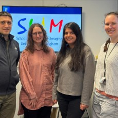
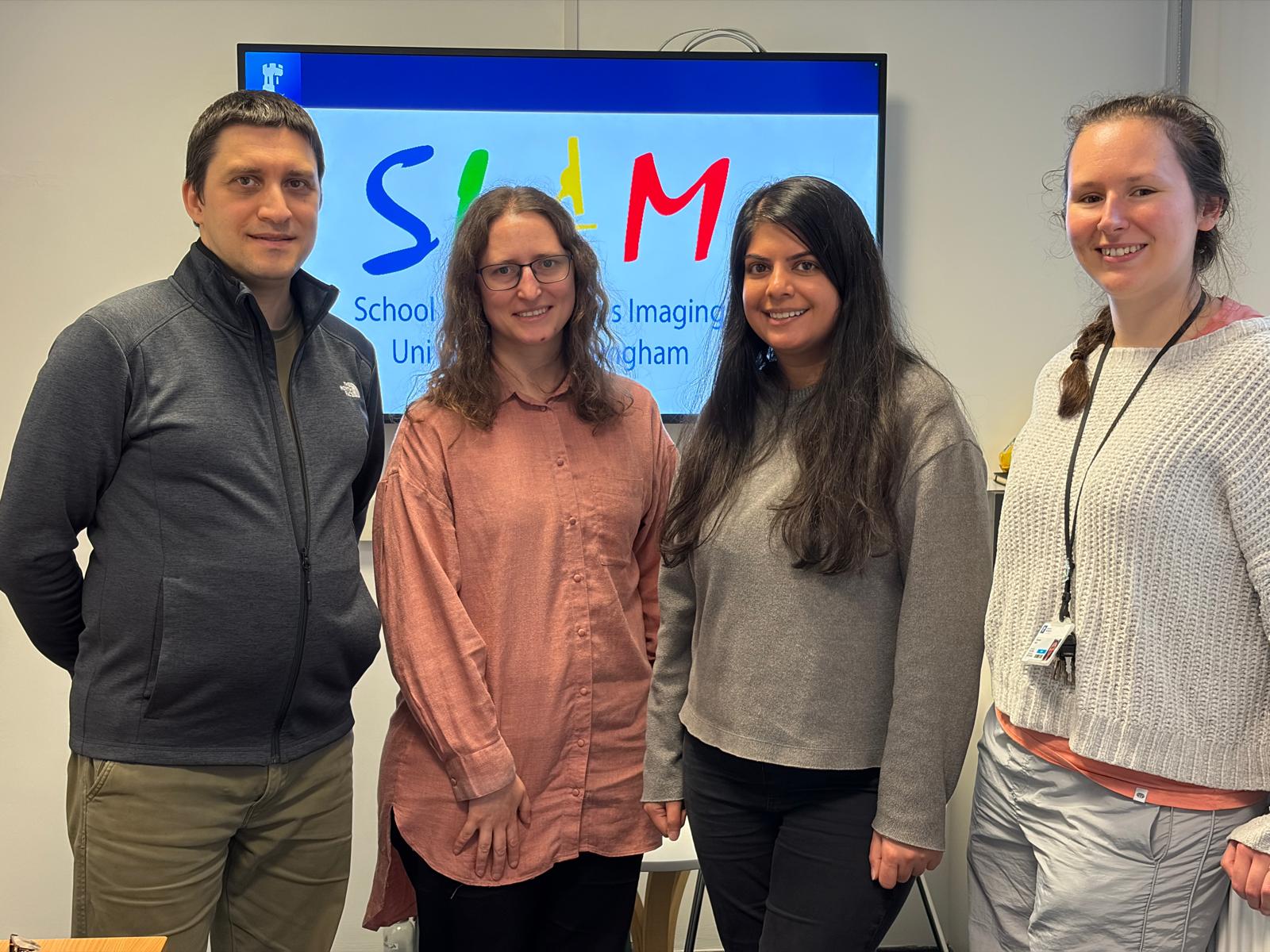
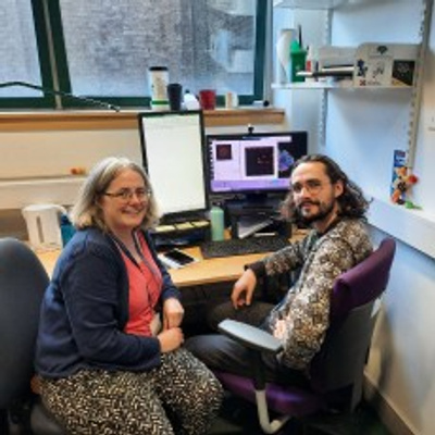
Spending a couple of days at the University of Nottingham shadowing Robert Markus and Seema Bagia at School of Life Sciences Imaging (SLIM) was a great experience. It was time to talk about good practice and processes, as well as get a sense of the ethos and working environment. Seeing how the team interacted with users and the wider research community whether in training or passing in the corridor made being there in person more valuable.
I spent time with the team seeing how they manage user access and bookings. Seeing the data management solution in place at SLIM from both the facility and user perspective that gave me lots of ideas for a system to implement at Leeds.
Robert showed me how he developed super-resolution imaging protocols for more challenging samples. I really appreciated having time to meet with one of the facility users to hear their experience of working with the facility - being trained, troubleshooting and gaining knowledge and confidence by working alongside the SLIM team. It highlighted to me the benefit of committing time to meet with users and their supervisors, particularly using more challenging techniques such as super-resolution, to support the image acquisition but also data processing and interpretation. It was clear that time with both users and their supervisors was helpful in building good working relationships with good understanding of the projects.
Thank you to Robert, Seema and the team for being great hosts, and to the RMS for making the trip possible.
My main outcome was to build in more time or better practices to enable conversations and more follow up with users to help them get the most from their projects. Having capacity for this may be challenging, but the value of taking time to help users understand their data was clear in my visit to SLIM.
The imaging Facility at Leeds is about to relocate, and I am hoping to use what I learned about the data management processes at SLIM to help inform the IT set up in the new space to best serve and support users and their data handling.
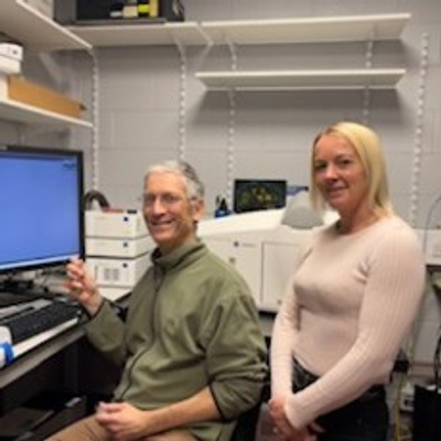
I visited the Flow Cytometry and Imaging facility in University of York. We spent time learning startup/shutdown procedures and how to operate cell sorters/analysers properly, as well as diagnosing common issues. They taught me about the theory behind flow cytometry measurements and the importance/purpose of wavelength compensation, and they let me practice using fluorescent beads. I also spent time with an engineer, troubleshooting/fixing a leaky flow cytometer. I had the chance to join the electron microscopy group, who were working on a new method to fix and analyse cells grown in a plastic multi-well plate. They demonstrated sample preparation, how light/electron microscopes work, what limits resolution, and the how to improve image quality. On top, I spent time with the microscopy group, learning about the theory and advantages of DNA-PAINT for improving resolution.
I've implemented new QC methods in our flow-cytometry lab. I've also learned how to operate a cell sorter, and how to tweak setting depending on cell types, which has inspired me to fix our cell sorter--it will be useful for several students in our molecular biology department, who would like to enrich and/or isolate specific cell types. I've also been teaching flow cytometry to other lab users, so they can collect data for their research.