Below you can find out the work that is being undertaken by our current RMS Diploma students:
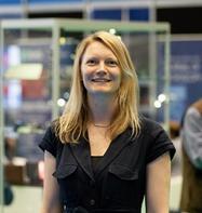
Developing protocols and training materials for the live imaging of Drosophila via lightsheet and multiphoton microscopy
Jennifer Adcott, University of Manchester, UK
Lightsheet and multiphoton microscopy have been widely used to reduce photo-damage. Here these systems will be used and compared for the imaging of live Drosophila using established cancer model fly lines. A protocol will be established for sample preparation, image acquisition, through to image processing and analysis. Suitability for each technique will be compared for addressing specific questions where live imaging of small model organisms or ex vivo tissues is required. The aim will be to publish these results as a methodology paper with JoVE video protocol.
The Characterisation of Speciality Fibres Utilising 3-D SEM Constructed Images
Michael Brookes, University of Leeds, UK
Distinguishing expensive speciality animal fibres such as cashmere and alpaca from cheaper fibres such as wool is an important activity. Current techniques, such as ISO 17751-2, measure fibre diameter utilising an SEM in 2 dimensions at high vacuum and susceptible to viewing errors. The study will use 3-D imaging techniques to measure and characterise speciality fibres and compare the results with that of current techniques.

Optimization of the preparation and analysis of Cementitious materials for Scanning Electron Microscopy
Stuart King, University of Leeds, UK
Scanning electron microscopy plays a major part in investigations of the microstructure of cementitious materials. The preparation, imaging and analysis of materials can be challenging due to the differences between the many types of cementitious materials. With the ongoing evolution in understanding these materials, the purpose of the study is to look at ways of developing the processes to optimize procedures in research.
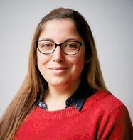
Evaluation of biological sample staining techniques using Uranyl Acetate and UA free options for TEM and a comparison of SEM chemical fixation techniques with Cryo-SEM
Silvia Martinez Micol, University of Bath, UK
This study explores the comparative effectiveness of Uranyl Acetate (UA) and UA-free alternatives in sample staining protocols for Transmission Electron Microscopy (TEM). It will evaluate different staining / fixation methods to enhance ultrastructural preservation and contrast while addressing safety and regulatory concerns associated with UA.
Key objectives include: Evaluating UA vs UA-free staining; Optimizing fixation protocols for reproducibility and image clarity; Assessing embedding and sectioning techniques (microtomy) for sample integrity. The study will also contrast chemical fixation methods with Cryo-electron microscopy (Cryo-EM), which preserves samples in a near-native hydrated state without chemical alteration.
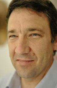
Thermally sprayed carbide composite microstructural characterisation by SEM, EDS and EBSD
Steven Matthews, University of Auckland, New Zealand
The wear resistance of carbide composite thermally sprayed coatings is critically dependent upon their microstructure, which is typically analysed via scanning electron microscopy using backscatter electron imaging, energy dispersive spectroscopy and electron backscatter diffraction. However, little has been presented on the effect of imaging conditions on the depth of BSE or EDS analysis, which is particularly challenging given the dramatic variation in material properties. This work aims to optimise sample preparation for SEM and quantify the depth of BSE/EDS analysis as a function of imaging conditions across composite phase interfaces from both the surface and the cross-section using FIB-capable SEM.
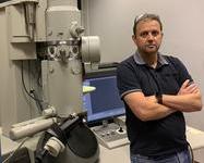
Accurate and precise particle size measurement of titanium dioxide using electron microscopy
Barry Millson, Tronox Pigments UK Ltd
From a regulatory perspective, the accurate and precise primary particle size measurement of particulate solids is becoming increasingly important since this defines which materials classified as a nanomaterial according to the revised EU definition (2022). For titanium dioxide producers, the particle size also affects the light scattering and hence, the optical performance of the product (in particular, opacity and tint tone). The project will focus on sample preparation methodology and measurement which is critical to increase the accuracy and repeatability of the particle size result of titanium dioxide. The study will also serve to increase my skills and knowledge in this important area.
Application of Deep Learning Tools for Imaging Cytometry to Enable Label Free Characterisation of Manufactured Red Blood Cells
Pamela Twist, Safi Biotherapeutics, UK
In the manufacture of red blood cells (RBCs), it is crucial to track the maturation of cells in the bioreactor from a hematopoietic progenitor cell to a mature RBC. This can be achieved by fluorescent staining of maturation markers. Using imaging cytometry, we can collect the fluorescent signal of cells along with their corresponding brightfield image that captures morphological changes of maturing RBCs. My study aims to investigate if we can apply deep learning tools to our imaging cytometry data, so we can characterise the stage of maturation of manufactured RBCs in a label free manner.

Rapid and Non-Destructive Analysis of fresh or Formalin-Fixed Tumor Biopsies Using Fluorescence Microscopy and Artificial Intelligence for Image Reconstruction
Krunoslav Vinicki, Digicyte Ltd, Croatia
The study aims to develop and validate a new approach in microscopy for the rapid and
non-destructive analysis of fresh tumor biopsy samples. With this method I'm planning to address a longstanding need in pathology for real-time, on-site biopsy examination while preserving the tissue for further analysis. This could expedite the initiation of targeted therapy and reduce the need for repeat sampling due to insufficient material.
Development of image analysis routines to study the dynamics of centrosomes and centrioles in Drosophila
Alan Wainman, University of Oxford, UK
Imaging has evolved significantly in the last few years, and no longer involves just the capture of beautiful images, but also the generation of large datasets that must be quantified and analysed. The main goal of this proposal is to develop my image analysis skills, initially through the attendance of courses and subsequently through the development of scripts for ongoing research. These skills will ultimately benefit not only my own research but also the support I provide to users of my imaging facility. I also hope to record my learning experience to help others hoping to develop their own image analysis skills.
Quantification and localisation of CD8+ T cells in the TME of Hh inhibitor treated mice by Light Sheet Fluorescence Microscopy
Timothy Young, University of Cambridge, UK
Cytotoxic (CD8+) T Cells are effective at eliminating cancer cells and their presence in the tumour microenvironment (TME) is associated with good prognosis in almost all cancer types. The de la Roche laboratory has found that CD8 infiltration into the TME is regulated by Hedgehog (Hh) signalling. When the key Hh signal transducer Smoothened (Smo) is knocked out or inhibited in CD8 T cells in mice, there is a greatly diminished anti-tumour response, resulting in increased tumour size. Specifically, pharmacological Hh inhibition in CD8 T cells resulted in reduced infiltration into the tumour mass. Although single plane imaging was performed, the full 3-dimensional overview of T cell infiltration in these tumours is unknown. Using Light Sheet Fluorescence Microscopy (LSFM), I aim to quantify and localise the extent of tumour infiltration of CD8+ T cells in Hh inhibited and vehicle treated mice.
