Browse through articles previously published in infocus Magazine. If you are an RMS member you can view any article. Non members will be able to read articles over a year old.
As an international society, the RMS has always sought to strengthen links with other organisations dedicated to furthering the science of microscopy around the world.
The RMS is very pleased to announce the winners of its latest awards, recognising scientific achievement, contributions to education and outreach, technical support and services to the Society.
Personally, I like recording videos of interesting or unusual things under the microscope to send to friends and family, or to include in publications.
We are moving into a new era in microscopy technology development in which the microscopy technologies, in addition to the imaging function they have always provided, increasingly provide us with additional predictive and prognostic information which would be impossible to derive by any other method.
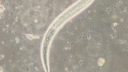
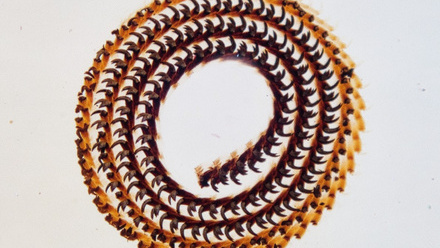
Our recently acquired collection of antique microscope slides includes some in the category “marine zoology”. The earliest members of our society – mainly wealthy, professional gentlemen – were keen to explore the new world that was opening up under their microscopes, and having neither the time nor skill to make their own, were avid collectors of slides made and sold by well-known mounters.
Formed in 1988, the Flow Cytometry Section is made up of a cross section of cytometrists representing research, clinical and industrial aspects of flow cytometry.
The scientific community increasingly recognises that laboratory practices can significantly contribute to the environmental footprint. At AstraZeneca, our ambitious sustainability strategy extends beyond corporate policies to fundamentally transform how we conduct research across our organisation and supply chain.
Microscopic meteorites is an art exhibition recently created by Dr Luke Norman and myself, to show the public the wonders of meteorites at the microscale, as well as (hopefully) educating them about space and space rocks at the same time.
Abstract: The acquisition of three-dimensional (3D) stereo paired images by scanning electron microscopy (SEM) typically requires the tilting of the SEM stage about the eucentric working distance, which is typically set at 10 mm working distance (WD).
The Journal of Microscopy, published by Wiley on behalf of the Royal Microscopical Society, is the world’s oldest journal dedicated to the science of microscopy. The Journal is critical to the RMS, as almost all the income it generates comes back into the society to help support our activities.
The surface characterisation of pharmaceutical-based powders is a challenge due to their beam sensitivity. Secondary electron hyperspectral imaging (SEHI) is a novel surface analysis technique which filters secondary electrons (SE) energies using mirror electrodes to generate a spectrum.
The aim of the studentship evolved significantly over the course of my placement with the Advanced Imaging Core Technologies Unit (AICTU). The central aim, however, of advancing my imaging skills across a range of microscopical and analytical techniques, remained consistent.
This RMS studentship was focused on creating and then testing the functionality of an optics module that would use the Raspberry Pi High Quality Camera modules instead of the currently used Raspberry Pi Camera 2 in the OpenFlexure microscope.