Enhancing your Image Course
Enhancing your Image Course
This is the second RMS workshop of this nature. This course aims to give delegates an introduction to the basics in colouring and enhancing images taken on a microscope with Adobe Photoshop, enabling them to make their images even more beautiful and inspiring. It is especially useful for colouring SEM micrographs and is designed to accommodate total beginners, with plenty of hands-on practice. Delegates will be asked to bring some of their own images to colour.
Photoshop-enhanced images are widely used in scientific publications, marketing materials and posters making the experience gained on this course invaluable and easy to implement back in the workplace. Coloured images also create more interest in the general public which helps science reach out to non-scientists.
Provisional Programme
Speakers
Scientific Organiser
-

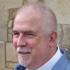
Ms Kim Findlay, FRMS
John Innes Centre
Kim is head of Bioimaging at the John Innes Centre, Norwich. Her degree was in Biology and Physics at King’s College, London. With over 34 years' experience in light and electron microscopy and more than 90 publications involving the use of TEM, SEM or confocal microscopy, in 2015 she was awarded the RMS Vice Presidents Medal for microscopy research and laboratory support. Her early focus on the plant cytoskeleton developed into a wider interest in plant and microbial sciences. She has made particularly important and long-standing contributions to Streptomyces research. Kim teaches cryo-SEM at the RMS EM school and taught on the RMS cryo-EM course in the past. She is regularly involved in Outreach activities, running tours and demonstrations for the public and young students. Kim won an award from the University of East Anglia, where she is an honorary lecturer, for her outstanding contribution to public and community engagement.
Speaker / Trainer
-
Mr Steve Gschmeissner
Photoquest
Steve Gschmeissner is one of the leading scanning electron microscopists in the world today. In the late 1990s he taught himself to colour using Photoshop and subsequently created an extraordinary collection of over 3000 pictures, covering specimens ranging from diatoms to cancer cells to new materials. His work is published globally in every conceivable type of media, including books, magazines, TV, advertising, record covers and most famously adapted by Damien Hirst in his biopsy series on the Cell.
Steve began his career in the more traditional role of an electron microscopist at the Royal College of Surgeons and later at UK Cancer Research. His training in the scientific environment informs his work with a deep understanding of the science behind the image. This knowledge, combined with a strong visual awareness of what makes an outstanding, oftentimes breath taking, image has won him numerous prestigious awards from the Royal Photographic Society, The Royal Microscopical Society and Spot on Micro.
Awards aside, the breadth of Steve’s collection has given us a glimpse into an extraordinary and magical micro-world of great curiosity and beauty.
Sponsors
-

FEI
See beyond with FEI's leading edge SEM, TEM, ESEM and DualBeam™ solutions. Our 60 year history of pushing the boundaries of microscope innovation has resulted in instruments delivering sub-nm SEM and sub-Å TEM resolution. Whatever your application in materials or life sciences, FEI delivers the highest performance solution, and puts you at the center of a global community of leading researchers and scientists.
Find out more about FEI
fei.com -

ZEISS
Carl Zeiss is an innovative technology leader in the fields of optics, precision engineering and electronic visualisation. Time and time again, we set new, pioneering standards in sophisticated technology for recognising, experiencing, measuring, analysing, structuring and processing a wide spectrum of objects. With professional optics we meet the expectations of even our most critical customers - not only in the fields of research, medicine, industry, but also for use in leisure activities.
Find out more about ZEISS
www.zeiss.com