
Australia Hosted Event
24 August 2026
14:00 - 15:00 AEST (06:00 - 07:00 BST)
Online
The expansion microscopy user group brings together researchers to share experiences to enable rapid uptake of the technology.
At this meeting... We will be joined by Dr Dawson Ling who will present his talk 'Ultrastructure Expansion Microscopy of the Malaria Parasite Tight Junction During Invasion'.
In this talk, Dr. Dawson will discuss his application of U-ExM to blood-stage Plasmodium falciparum malaria. The invasion of red blood cells requires precise coordination of specialised parasite organelles, cytoskeletal structures and host–parasite interactions that occur at the nanometre scale. He will describe how expansion microscopy provides new opportunities to resolve parasite ultrastructure and investigate the organisation of invasion machinery beyond the capabilities of conventional microscopy.
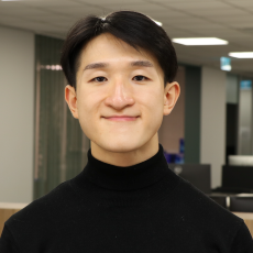
Dawson Ling is a postdoctoral researcher in the Centre for Dynamic Imaging at WEHI. His research focuses on understanding how Plasmodium falciparum invades human red blood cells using advanced microscopy approaches, including lattice light sheet microscopy and expansion microscopy. He completed his PhD at the Burnet Institute, where he investigated the mechanism of action of invasion blocking antimalarial compounds and identified parasite proteins involved in invasion-related lipid metabolism and signalling.
Organisers:

Gabriela Segal Wasserman
Dr Segal has a PhD in Tumour Immunology from the Dept. of Microbiology and Immunology at the University of Melbourne. She has years of experience in sample preparation for confocal and multiphoton microscopy. In her current role as an academic specialist she supports confocal, tissue clearing and Lightsheet applications.
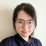
Wing Tung Wong
Wing Tung Wong is a PhD student at the University of Technology Sydney, specialising in expansion microscopy to achieve super-resolution imaging, focusing on developing an approach for internalised extracellular vesicles with different host cells, and spheroids interactions.
RMS Organisers:

Organised by:

