
Australia Hosted Event
3 December 2026
16:00 - 17:00 AEDT (05:00 - 06:00 GMT)
Online
The expansion microscopy user group brings together researchers to share experiences to enable rapid uptake of the technology.
At this meeting we'll be joined by Associate Professor Toby Bell who will present his talk 'Expansion Microscopy in the Cell Nucleus'.
In this talk Toby will present two of their recent applications of expansion microscopy in the cell nucleus. The first involves Hendra virus matrix protein which imaging reveals transits to sub-nucleolar compartments before relocating to the cell membrane and participating in budding. The second application is in the mapping of the distributions of histone post translational modifications in T cells. They show that the spatial arrangement of these is related to their roles in chromatin compaction and changes considerably as T cells undergo differentiation. Some recent work towards combining expansion microscopy with single molecule super-resolution will also be discussed.
Toby Bell is an Associate Professor in the School of Chemistry at Monash University where he heads a multidisciplinary research group which uses time- and space-resolved fluorescence techniques to investigate systems ranging from small molecules to whole cells. A particular focus of the group’s research over recent years has been the development and application of super resolution microscopy techniques to study cellular ultrastructure, organelles and the cell nucleus. Current research areas include how viruses evade cellular defence mechanisms, how the spatial arrangement of epigenetic modifications in T cells regulates differentiation and immunological memory, and the development of new fluorescent dyes and nanoparticles for imaging, sensing and diagnostic applications. Toby received his PhD in 2002 from the University of Melbourne during which he studied energy and electron transfer phenomena in porphyrin-based donor-acceptor compounds. This was followed by postdoc stays firstly at the Max Planck Institute for Radiation Chemistry in Germany and then at the University of Leuven in Belgium where he applied single molecule spectroscopy to photoactive dendritic and polymeric materials. Toby returned to Australia in 2006 as a Centenary Research Fellow at the University of Melbourne before commencing at Monash University in 2009.
Organisers:

Gabriela Segal Wasserman
Dr Segal has a PhD in Tumour Immunology from the Dept. of Microbiology and Immunology at the University of Melbourne. She has years of experience in sample preparation for confocal and multiphoton microscopy. In her current role as an academic specialist she supports confocal, tissue clearing and Lightsheet applications.
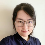
Wing Tung Wong
Wing Tung Wong is a PhD student at the University of Technology Sydney, specialising in expansion microscopy to achieve super-resolution imaging, focusing on developing an approach for internalised extracellular vesicles with different host cells, and spheroids interactions.
RMS Organisers:

Organised by:

