RMS Early Career Award
The annual Early Career Award recognises the achievements of an outstanding early career imaging scientist in their contribution to microscopy, image analysis, or cytometry.
This contribution may be through an impressive research application, development of techniques or analysis tools, an inspiring public engagement initiative, or a demonstration of exceptional support to other members of the community.
Applications for the 2026 Early Career Award have now closed.
The 2027 Early Career Award application window will open in early 2027.
How to submit a nomination:
Applicants should submit the following two documents:
- An Applicant Statement which describes the following (max. 300 words):
- The merits of the Applicant
- A brief summary of the Applicant's work, accompanied by no more than one figure (sub panels acceptable within reason).
- A statement of the Applicant's suitability for the award
- Please state your career stage/time in microscopy field at the start of your application (this does not contribute to the 300-word limit).
- A Letter of Support from their Line Manager/Supervisor covering the following (max. one-page):
- The novelty of the contribution in the context of the prior state of the field.
- The impact of the contribution.
- The applicant’s role in leading the delivery of the contribution.
Eligibility:
Any person undertaking work in the field of microscopy/image analysis/flow cytometry and belonging to one of the following categories is eligible for this award*:
- Current undergraduate/postgraduate/Masters/PhD students; or
- Within 8 years of starting work or studies in a microscopy-related field (excluding career gaps)
All applications received will be reviewed by members of the RMS Early Career Committee and three finalists will be shortlisted to give an in-person 5-minute flash talk at: Microscopy: Advances, Innovation, Impact 2026 meeting, incorporating the RMS AGM on Wednesday 30 September 2026 at the Royal Society of Chemistry, London.
The three shortlisted finalists will be able to claim up to £200 towards their travel/accommodation expenses to attend the meeting.
In addition to the above you may be eligible to apply for an RMS Bursary for further financial support to attend the meeting. You can find further information about RMS Bursaries on our website.
Additional Statement:
We have now implemented an Additional Statement option for the award. This is completely optional. If you feel you have faced significant personal, societal or institutional barriers to your achievements and would like to disclose this, in addition to the two documents above please send an Additional Statement (max 50 words). This will be kept confidential from the Early Career Committee and will only be reviewed by a pre-appointed member of RMS staff to fairly weight your final score.
We are trialling the Additional Statement option for the first time, and we hope that it will make our award more inclusive. We have spent time thinking about how best to run this option but as it is our first year, please bear with us!
Judging criteria:
Impact of the work (10 points)
Impact can include an impressive research application, development of techniques or analysis tools, an inspiring public engagement initiative, or a demonstration of exceptional support to other members of the community. This list is non-exhaustive.
Novelty of research or innovation of stewardship (15 points)
The uniqueness of the research/contribution, extent to which it builds on existing practises/research, the originality of thinking, and the applicant’s role in its delivery. Set within the career stage of the entrant and the extent to which the microscopy community is part of this, whether this is the microscopy community or a development of a microscopical technique.
Presentation and accessibility of application (5 points)
Ease of understanding of the application to a non-specialist. Is the entry engaging and can it stand alone to demonstrate the entrants merits.
The 2026 award-winner will receive the RMS Early Career Award and a £100 cash prize.
2025 Winner
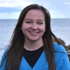
Niamh Burke, University College Dublin
Niamh won this year's award during the Early Career Symposium at mmc2025 in early July, where she gave a talk about her work on the enderscope community project - co-developing accessible imaging tools with the public.
Niamh is a final-year PhD student and Anatomy Demonstrator at University College Dublin (UCD) and a Postgraduate Representative with the Microscopy Society of Ireland. During her undergraduate degree in Physiology in UCD, Niamh completed a short project under the supervision of Dr Mark Pickering. It was here where she developed a strong interest in building simple, low-cost, open-source lab instruments. Taking a brief break from the world of open hardware, Niamh then worked as a Research Assistant in the Cancer Biology and Therapeutics Group in the UCD Conway Institute under Professor William Gallagher. Following this, Niamh returned to the Pickering Lab for her PhD, which focuses on designing open and accessible tools to measure microplastic pollution in the sea.
As the representative for the Microscopical Society of Ireland on the RMS Early Career Committee, Niamh works to increase engagement and encourage networking between the RMS and Irish Early Career Researchers.
2024 Winner

Akaash Kumar, MRC Laboratory of Molecular Biology
Akaash was awarded the RMS Early Career Award for his work developing multispectral live cell imaging which allows for up to six fluorophores to be imaged simultaneously
Akaash is a recent Post-Doctoral Scientist in the Cell Biology Division at the MRC Laboratory of Molecular Biology. A biochemical engineer by trade, he is interested in developing technology to help solve biological problems, often relying on interdisciplinary expertise from engineering and physics. As part of his PhD, at the University of Cambridge and the MRC Laboratory of Molecular Biology, he developed novel multispectral, video-rate fluorescence microscopy hardware and software which can improve any camera-based fluorescence microscope globally. He is applying this technology to study the mechanisms of endosomal sorting and trafficking more broadly within mammalian cells.
Akaash has been awarded a PhD Award by the Cambridge Society for the Application of Research in recognition for the impact of his PhD work and he has presented his research both nationally and internationally, having recently won Best Talk Awards at the Warwick-Cambridge Quantitative Cell Biology Symposium 2024 and RMS Frontiers in Bioimaging 2022. He is also an inventor on the patent application describing his PhD work and the technology is currently undergoing commercialisation with a major microscopy company.
Prior to his PhD, Akaash worked at the Wallace H. Coulter Dept. of Biomedical Engineering at Georgia Institute of Technology and Emory University. Whilst there he developed novel bioprocessing technology for stem cell manufacturing and had a significant role in undergraduate/graduate-level teaching. He has also received an MSc in Advanced Chemical Engineering with Biotechnology from Imperial College London and a BEng in Biochemical Engineering from University College London.
2023 Winner
Alex Johnson, Medical University of Vienna
Alex was awarded the RMS Early Career Award for his work developing a range of quantitative light and electron microscopy approaches to investigate the molecular mechanisms of endocytosis in plants.
He started working with plants during his 1st post doc in the labs of Dr. Gregory Vert and Dr. Christien Merrifield (Centre National de la Recherche Scientifique, France). At this time, how endocytosis functioned in plants was poorly characterized. This is because plants present many technical challenges to image dynamic sub-cellular processes, like endocytosis, at appropriate resolutions. However, during this placement, Alex and his colleagues established a reliable method to apply total internal reflective fluorescence microscopy (TIRF-M) with automated unbiased analysis to quantify live single events of endocytosis in intact plants. Building on this experience, he then joined the group of Prof. Jiri Friml (Institute of Science and Technology Austria, Austria) where he optimized the use of structured illumination microscopy (SIM) to further improve their ability to resolve the dynamic interplay of proteins at endocytic events. Furthermore, here he also developed the use of metal replicas of unroofed plant cells with scanning electron microscopy (SEM) and scanning transmission electron microscopy (STEM) tomography to directly visualize plant endocytosis events in 3D for the first time. Using these quantitative imaging tools, Alex has significantly updated the working model of plant endocytosis and uncovered that it is evolutionarily unique from other eukaryotic model systems. In addition, Alex has released details of the the full protocols, analysis codes and example data for these microscopy and analysis techniques as open access resources to aid the progression of plant endocytic investigation.
He is currently a post doc in the lab of Dr. Kareem Elsayad (Medical University of Vienna, Austria), where he is developing novel optical microscopy and spectroscopy techniques to elucidate microscopic scale mechanical, structural, and dynamic properties of biological samples.
2022 Winner

Katherine Paine, University of York
Katherine, who began her PhD in 2018 at Chris MacDonald’s laboratory, was chosen in recognition of the novel approaches in imaging and cytometry she has brought to her studies on the regulation of cell surface membrane proteins.
Cell surface membrane proteins perform diverse and critical functions and are spatially and temporally regulated by membrane trafficking pathways. These trafficking pathways are evolutionary conserved from yeast to humans. MacDonald lab uses yeast as a model organism to study these pathways.
It became clear from Katherine’s initial studies that although standard confocal microscopy could be used to visualise some of the processes she was interested in, there were also limitations. She then helped optimise a suite of imaging and cytometry approaches to study surface proteins. This includes Airyscan2, structured illumination (SIM) and photoactivated localisation microscopy (PALM); all of which can be coupled to bespoke microfluidic exchange systems.
Katherine is also in the process of optimising a high throughput method to measure Förster resonance energy transfer (FRET) in yeast using robotics and flow cytometry.