Taking place every two years, this exciting international competition is a great opportunity for microscopists from across the world to showcase their skills. It provides a great outlet for those that can capture the breath-taking and engaging beauty of the microscopical world. The Society is always in awe of the quality of submissions across the categories.
The RMS Scientific Imaging Competition 2025 took place as part of mmc2025 incorporating EMAG 2025.
2025 Winners
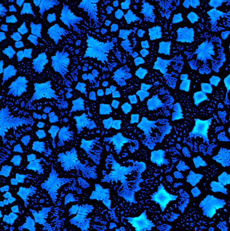
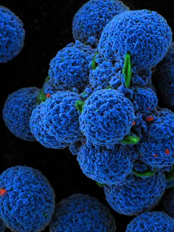
Peptide snowflakes
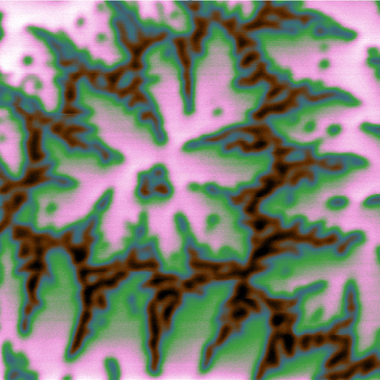
Magnetic flowers
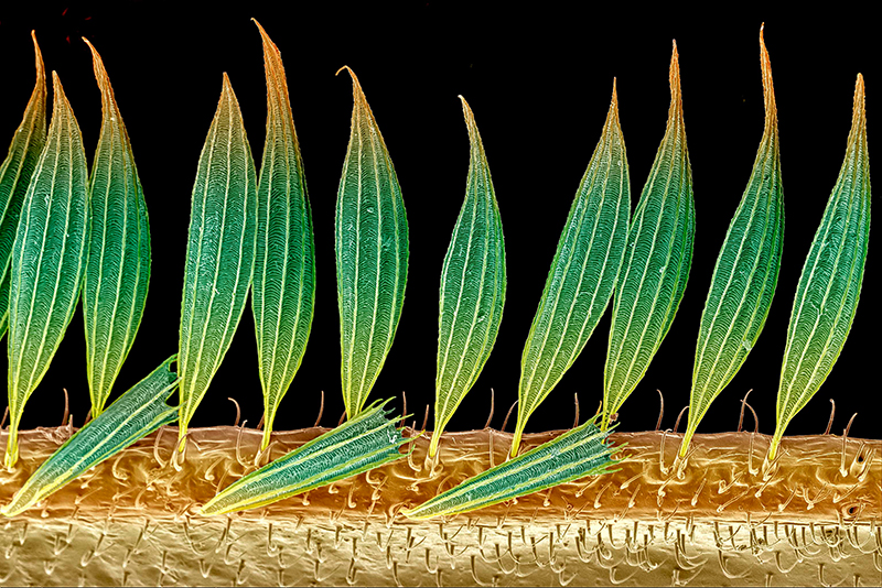
"Feather-like" Structures
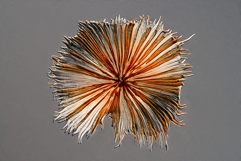
Flowering heads of aosa rupestris
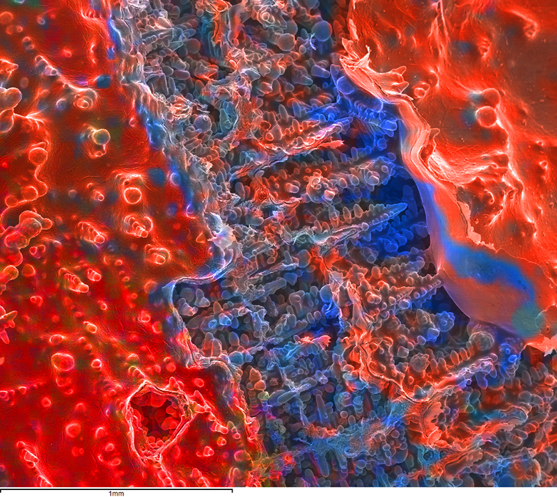
NMC particles with Zr
Ductile iron lustrous carbon defect
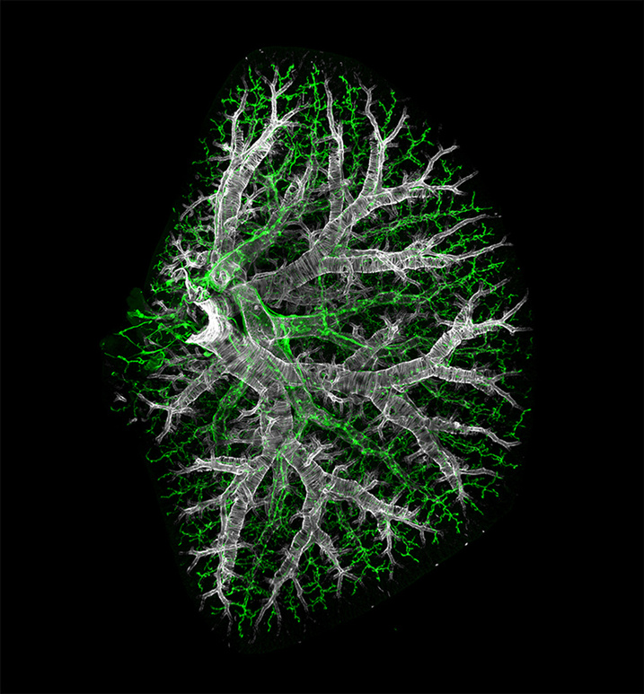
Lymphatic vessels and smooth muscle in a cleared lung
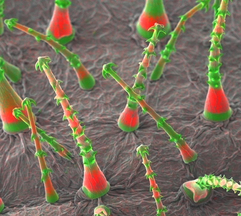
Trichome
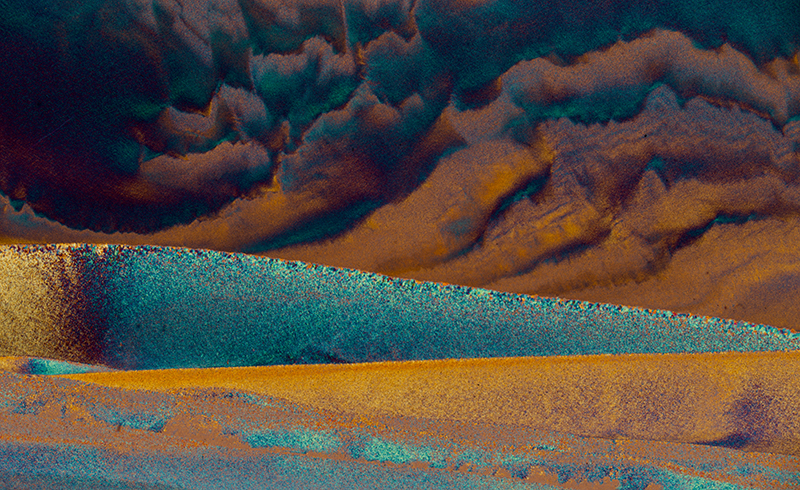
Storm approaching
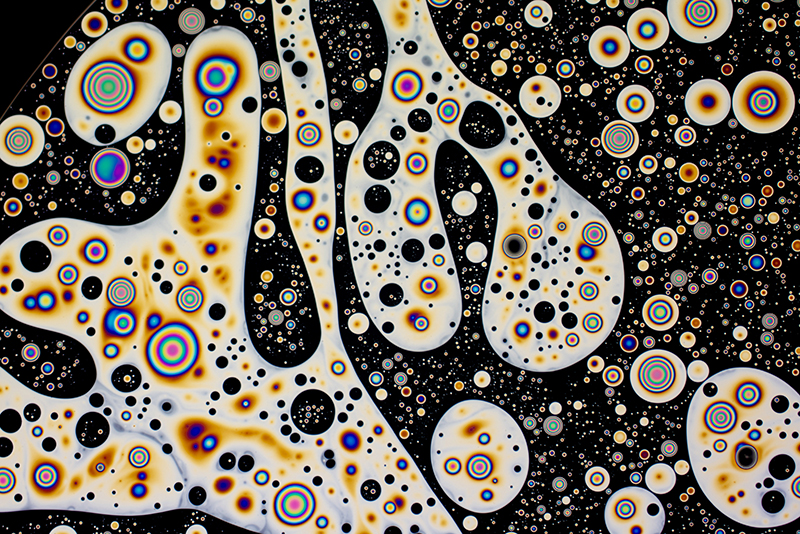
Colourful Micelles